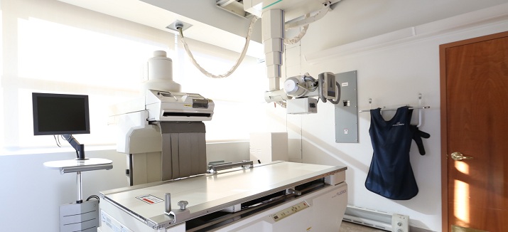
Fluoroscopy is a special x-ray technique that makes it possible to see internal organs in motion. Through a series of consecutive x-rays, a video of the organ of interest is generated.
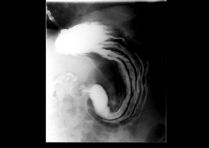 Esophogram, Upper GI, and Small Bowel Series are types of x-ray examinations of the esophagus, stomach, and small intestine respectively.
Images are produced using x-rays, fluoroscopy and liquid barium contrast. Through a series of consecutive x-rays, a video of the organ of interest is generated.
Patients drink barium to coat the inside of the GI tract making it much more visible to x-ray and fluoroscopy.
Do not eat or drink anything after midnight on the day of the examination. On the day of the exam, please wear loose, comfortable clothing, avoiding garments that have zippers, belts, or buttons made of metal. You may be asked to wear a gown during the procedure. You will be asked to remove keys and jewelry in the area being scanned.Women should always inform their physicians and the technologist if there is any possibility of pregnancy.
You will be comfortably positioned by a technologist either in a standing or lying down position. As you drink the liquid barium, our radiologist will watch the barium pass through your digestive tract. You will be positioned at different angles to take several x-ray images. After the necessary images are obtained, the examination is complete. The examination is typically completed in about 15-20 minutes for esophograms and upper GI series. For small bowel series, the examination can range from one hour to several hours.
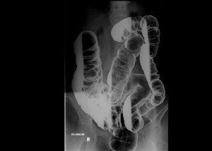 A barium enema is an x-ray examination of the colon.
Images are produced using x-rays, fluoroscopy and liquid barium contrast. Through a series of consecutive x-rays, a video of the colon is generated.
Barium is used to coat the inside of the colon, making it much more visible to x-ray and fluoroscopy.
For the 24 hours prior to the exam, do not eat solid food. Drink only clear liquids like juice, tea, black coffee, cola or broth, and avoid dairy. After midnight the night before the exam, do not eat or drink anything. You will take a laxative medicine the night before the exam and a cleansing enema in the morning a few hours before your appointment.On the day of the exam, please wear loose, comfortable clothing, avoiding garments that have zippers, belts, or buttons made of metal. You may be asked to wear a gown during the procedure. You will be asked to remove keys and jewelry in the area being scanned.
Women should always inform their physicians and the technologist if there is any possibility of pregnancy.
You will be comfortably positioned on the examination table by a technologist. A small tube will be inserted into your rectum and will be used to coat your colon with barium. Air may also be injected through the tube to help the barium thoroughly coat your colon. You will be frequently repositioned in order to see your colon from several angles. After the necessary images are obtained, the examination is complete. The examination is typically completed in about 30 minutes.
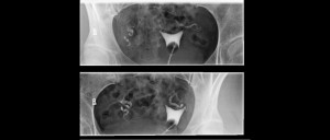 A hysterosalpingogram is an x-ray examination of a woman’s uterus and fallopian tubes. Images are produced using x-rays, fluoroscopy and liquid contrast dye. A series of low dose x-rays will be necessary to produce a picture of the organ of interest in motion. During a hysterosalpingogram, the uterus and fallopian tubes are filled with a contrast dye allowing the radiologist to view and evaluate their anatomy and function using fluoroscopy.
The hysterosalpingogram is best performed one week after menstruation but before ovulation to make certain that you are not pregnant during the exam. Our office can accommodate you to fit your scheduling requests.
On the day of the exam, please wear loose and comfortable clothing. You will be asked to wear a gown during the procedure. Prior to the procedure, you may be given a mild over-the-counter medication to minimize any potential discomfort.Women should always inform their physicians and the technologist if there is any possibility of pregnancy.
The procedure is like a gynecological exam with you positioned on your back on the exam table and knees bent. A speculum is then inserted which allows for a catheter (thin tube) to be inserted into the cervix. The speculum is removed and you are repositioned underneath the fluoroscopy camera. The contrast material then fills the uterine cavity, fallopian tubes and peritoneal cavity through the catheter.Our radiologist will monitor in real-time your images on a computer screen next to you as the exam is being performed. You will be frequently repositioned in order to see your uterus and fallopian tubes from several angles. After the necessary images are obtained, the examination is complete. The examination is typically completed in about 30 minutes.
