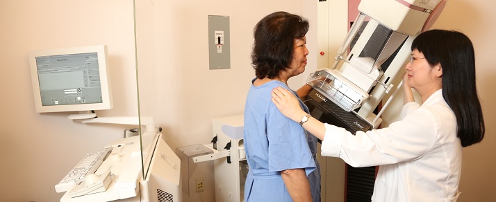
CP Advanced Imaging understands the importance of early detection and preventive care. We want to inspire women to be proactive about their health. Our women’s imaging services specialize in breast imaging, imaging of the pelvis and reproductive organs and diagnosing osteoporosis. Our caring staff and on-site radiologists are committed to providing exceptional care to our patients.
With digital mammography, ultrasound and MRI, CP Advanced Imaging is committed to providing women with the best and most comprehensive evaluation of the breast.– Digital Mammography is a low-dose x-ray examination of the breast used in the early detection and diagnosis of breast diseases. Current national guidelines recommend annual screening mammography for all women age 40 and over.
– Breast tomosynthesis or 3D mammography is an FDA approved advanced technology that can improve detection of breast cancer, particularly in women with dense breast since it provides a clearer image of dense breast tissue.
– Breast Ultrasound uses high-frequency sound waves to produce images of the breast. Ultrasound does not use ionizing radiation and does not involve x-ray exposure. It is commonly performed together with mammogram except in younger women where it can serve as the initial test. Breast ultrasound is also commonly used to evaluate women with dense breast tissue which is very common and is not abnormal. However, dense breast tissue can make it harder to find cancer on a mammogram and may also be associated with an increased risk of breast cancer.
– Breast MRI uses a magnetic field to provide highly detailed pictures of the breast to help evaluate a wide range of conditions. MRI does not use ionizing radiation and does not involve x-ray exposure. Breast MRI allows physicians to better determine the presence of certain diseases that may not be assessed adequately with other imaging methods such as mammogram or ultrasound.
With ultrasound and MRI, CP Advanced Imaging is committed to providing women with the best and most comprehensive evaluation of the uterus and ovaries.– Pelvic Ultrasound uses high-frequency sound waves to produce images of the uterus and ovaries and is most commonly the initial test and often times the only necessary test for evaluation.
– Pelvic MRI uses a magnetic field to provide highly detailed pictures of the pelvis to help evaluate a wide range of conditions such as pelvic pain, ovarian masses, fibroids and uterine abnormalities. The detailed MR images commonly allow physicians to better determine the presence of certain diseases that may not be assessed adequately with other imaging methods such as ultrasound.
– Sonohysterogram and Hysterosalpingogram are specialized tests to evaluate the uterus and fallopian tubes. These tests provide additional information to the conventional pelvic imaging tests.
CP Advanced Imaging understands the important role that bone quality factors into women’s health. Bone density scanning uses low dose x-ray to measure bone loss and to predict risk of bone fracture. Many national guidelines recommend testing all women age 65 and over as well as in postmenopausal women with risk factors for bone loss.


